Under-Eye Bags & Dark Circles · Newport News
Under-eye bags and dark circles in Newport News.
Dark circles have five different causes and filler only fixes one. We work out which you have first — on Jefferson Avenue near Kiln Creek — and then treat the one you actually have.
“Dark circles” is a symptom, not a diagnosis
Most clinics will offer you tear trough filler before establishing what is actually causing the problem. That works when the cause is a volume deficit at the rim. It does nothing when it is not — and most of the time, it is not.
The under-eye region has five distinct problems that all look similar in a bathroom mirror. They respond to completely different treatments. Getting the diagnosis right is the entire job.
The five causes
1 · Vascular — bluish, violet or purple
The skin of the lower eyelid is the thinnest on the body, roughly half a millimetre. Beneath it sits a dense venous plexus. Thin, translucent skin transmits that colour to the surface as blue-violet discoloration — the same optical effect that makes wrist veins look blue.
Tells: the colour reads bluish or purple rather than brown. It worsens with fatigue, dehydration, congestion and allergy season. Stretch the skin gently sideways with two fingers and it fades or blanches.
This is the cause most often written off as untreatable. Patients are told for years that their dark circles are “just genetic” and that nothing works. Skincare does not work on it, because there is nothing wrong with the skin. Filler does not work on it, because there is no volume deficit. But the vascular component responds directly to the right laser, and it is frequently the single largest contributor to how tired someone looks.
2 · Pigmentary — brown or tan
Melanin deposition in the periorbital skin. Often familial, more common in Fitzpatrick III–VI, and worsened by eczema, chronic rubbing, allergic shiners or prior inflammation.
Tells: brown rather than blue. Does not blanch on stretch. Frequently present in other family members.
This is the cause most easily made worse by aggressive treatment. Heat and inflammation in melanin-competent skin produce post-inflammatory hyperpigmentation. Treatment starts topical and stays conservative.
3 · Structural shadow — the tear trough
Not pigment at all. A groove runs from the inner corner of the eye down and laterally along the orbital rim, where skin is tethered to bone by the tear trough ligament. It deepens with age and casts a shadow.
Tells: the “darkness” changes dramatically with lighting. It nearly disappears under flat frontal light and looks worst under overhead light. Photograph yourself under a ceiling light, then facing a window, and compare.
Critically — and this is where most treatment plans go wrong — this groove is usually a downstream consequence of what is happening in the cheek, not a local problem in the lid. More on that below.
4 · Fat pseudoherniation — true under-eye bags
The orbital septum weakens and orbital fat bulges forward into the lower lid. A genuine bag: a convexity visible in profile, present on waking, not fluid, not shadow.
Tells: it protrudes. More visible in profile or three-quarter view. Does not change with hydration or sleep. May worsen on upgaze.
Nothing non-surgical fixes this. Not laser, not microneedling, not radiofrequency, not PRP. Fat that has herniated through a weakened septum has to be repositioned or removed, and that is a lower blepharoplasty.
5 · Textural — crepey skin, fine lines, laxity
Thin, wrinkled, lax lower lid skin from photodamage and elastosis. Can present as festoons — malar mounds of chronic lymphoedema below the orbital rim.
Tells: fine crosshatched lines, tissue-paper skin on animation, a distinct mound over the cheekbone that puffs in the morning.
Most people have two or three of these at once. That is why single-modality answers fail.
How we work it out
Your consultation is a physical examination, not a sales pitch.
Stretch test. Vascular discoloration blanches under lateral traction. Pigment does not.
Lighting assessment. Photographed under both flat and oblique light. Shadow-driven darkness changes between the two; pigment and vascularity do not.
Profile and dynamic views. Fat pseudoherniation is obvious in profile and on upgaze. Shadow is not.
Midface assessment. Where the lid-cheek junction sits, how much deep compartment volume has been lost, and whether the groove is primarily local or a consequence of midface descent.
Lid laxity testing. Snap-back and distraction. Poor tone changes what we are willing to do and how deep we are willing to go.
Dermatoscopy. Polarised dermatoscopy to separate epidermal from dermal pigment, and to clear any pigmented lesion in the field before a laser goes near it.
You leave with a written assessment of which causes you have, in what proportion, and what each one costs to treat — including the ones we cannot treat.
The treatments, and what each one is for
Long-pulse 1064 nm Nd:YAG — for vascular discoloration
A long-pulse Nd:YAG delivers energy preferentially absorbed by haemoglobin. Pulse duration is matched to the thermal relaxation time of the target vessel, so the vessel wall is heated and coagulated while surrounding tissue is spared. The vessel then clears over the following days to weeks. On our platform this is a small-spot vascular handpiece, which is what suits discrete vessels in thin lid skin.
This is the right tool for the blue-violet under-eye and for visibly prominent reticular veins in the lower lid and temple — a finding most patients have never been offered treatment for at all.
Session 15–30 minutes, with chilled air cooling throughout and topical anaesthetic available. Downtime mild redness and swelling for 24–72 hours, occasional bruising over larger vessels; most people work the next day. Timeline the fastest-responding treatment on this page — clearance is often visible within the first week. Course typically one to three sessions at 4–6 week intervals.
What it does not do: pigment, shadow or fat. If your discoloration is brown, or your problem is a bulge, this is the wrong laser and we will tell you.
Eye protection. Any treatment at or inside the orbital rim is performed with internal metal corneal shields. External goggles protect the provider, not the patient. Ask any clinic you are considering — including us — exactly what shielding they use.
Q-switched Nd:YAG — for pigment, selectively
Q-switched pulses in the nanosecond domain deliver energy faster than heat diffuses out of a melanosome, producing photomechanical fragmentation of pigment rather than bulk heating. At low fluence over multiple sessions this can lighten periorbital hyperpigmentation.
We use it selectively. Periorbital pigment in darker skin carries genuine risk in both directions — post-inflammatory hyperpigmentation and hypopigmentation. In most pigmentary cases we start with a topical regimen and reassess at twelve weeks before considering a device. If you have brown periorbital pigment and a clinic’s first move is a laser, get a second opinion.
Er:YAG resurfacing — for texture and fine lines
Erbium:YAG sits near the absorption peak of water, which makes it the most precisely controllable ablative wavelength available. Each pulse vaporises a thin, predictable layer with a narrow zone of residual thermal damage, and depth is set by the number of passes rather than by a preset.
Our laser has no fractional lens and we will not describe a capability we do not have. What we offer around the eyes is a light peel through full-field resurfacing, with the depth chosen against lid laxity, skin thickness and Fitzpatrick type. The periorbital region is one of the highest-value indications for erbium — thin skin that heals quickly, carrying a heavy wrinkle burden.
Session 30–60 minutes including 45–60 minutes of topical anaesthetic. Downtime four to seven days depending on depth: pinpoint bleeding, then bronzing, then flaking. Real social downtime — plan around it. Timeline texture at two to four weeks, collagen remodelling continuing to three to six months. Course one to three sessions.
Antiviral prophylaxis for every facial Er:YAG regardless of cold sore history. Corneal shields for anything at or inside the rim. Detail on the laser resurfacing page.
RF microneedling — for laxity and texture
Insulated needles deliver radiofrequency energy at a controlled depth in the dermis, creating discrete thermal coagulation zones that drive collagen remodelling. Because energy is delivered below the epidermis rather than through it, the epidermal risk profile is favourable across skin types — which makes it one of the few tightening technologies appropriate for darker skin.
Depth is the whole treatment. The infraorbital region, the malar region and the jawline are three completely different tissue thicknesses, and running one set of parameters across all of them is how people get hurt. We titrate needle depth and energy to the tissue we are in, and the settings used at the lid-cheek junction bear no resemblance to those used on the lower face. This is technique-dependent work, which is why it is not handed to someone trained on a single protocol. See RF microneedling.
Downtime two to four days of redness and grid-pattern swelling. Course typically three sessions at 4–6 week intervals, progressive over three to six months.
Microneedling, with or without PRP
Mechanical microneedling creates controlled micro-injury that stimulates collagen without thermal energy. Depth is set to lower lid skin thickness, which is a fraction of what is appropriate elsewhere on the face.
Platelet-rich plasma is prepared from your own blood — drawn, spun and separated in the same visit and in front of you. Applied during microneedling, it delivers concentrated growth factors into the channels the needles create. The preparation protocol is on our PRP page.
Honest positioning. The published evidence for PRP specifically around the eye is thin. The most-cited trial treated ten people in a single session without a control group, and found a statistically significant improvement in infraorbital colour homogeneity but no significant change in melanin content, skin hydration, wrinkle volume or wrinkle visibility. That is a real finding and a narrow one. We offer PRP here as an adjunct that may help colour, not as a treatment for texture or wrinkles, and anyone promising transformation from PRP alone is overselling it.
Downtime 24–48 hours of redness. Course three to four sessions at four-week intervals.
We no longer offer PRP injected on its own. Intradermal PRP across the infraorbital region was on this page as a separate treatment; it is not any more. The evidence that carried it was one uncontrolled ten-person trial, which is not enough to justify billing for a course of injections in an area where bruising is the norm. Where PRP earns its place around the eye, it earns it applied during microneedling, which is the version above.
Why we usually treat the cheek, not the tear trough
This is the part of the plan most clinics skip, and it is the reason a lot of people are sold filler they did not need.
The groove under your eye is usually not a hole that appeared in your lid. It is the edge of a shelf that dropped.
The tear trough and orbital retaining ligaments anchor skin to the orbital rim. They do not move. The deep fat compartments of the midface deflate and descend with age. When the tissue below a fixed anchor point falls away, the anchor point becomes a groove and a step-off appears at the lid-cheek junction. What you perceive as a hollow under your eye is frequently the shadow cast by a cheek that has lost support.
That has a direct treatment implication: rebuild the platform and the groove softens, without putting anything into the lower lid. A structural volumiser placed deep on the periosteum in the medial and lateral cheek restores the shelf, shortens the shadow and improves the lid-cheek transition. Temple and lateral cheek support add vector to the same effect.
It is further from the dangerous anatomy. The infraorbital region contains branches of the angular artery, which anastomoses with the ophthalmic artery. The tear trough appears in published series of filler-related visual loss. Deep cheek placement is well away from that anastomosis.
No Tyndall effect. Product placed deep under thick cheek tissue cannot scatter light through half-millimetre lid skin the way superficial lid product does.
No malar oedema. The deep cheek compartments have functional lymphatic drainage. The lower lid does not. Hyaluronic acid binds many times its weight in water, and in a lid with poor drainage that produces persistent puffiness that can last years and resists hyaluronidase.
It lasts longer, and it treats the cause. Structural product in a deep, low-mobility plane outlasts soft product in a high-mobility lid by a wide margin. The lid was not the problem. The cheek was. See dermal fillers and facial balancing.
Combine that with vascular laser to remove the colour and resurfacing or microneedling to thicken the envelope, and a substantial share of patients who walk in asking for tear trough filler no longer want it. Not because we talked them out of it — because the thing they were trying to fix is gone.
When direct tear trough treatment is genuinely the answer: an isolated structural groove at the rim, good lower lid tone, no fat pseudoherniation, no history of festoons or malar oedema, adequate skin thickness, and a midface that is already well supported. That patient exists. They are just far less common than the marketing suggests.
When it backfires: filling below a true fat bulge does not remove the bulge, it creates a longer, smoother convexity from lid to cheek. Filling a lid with festoons produces worse festoons. Filling a groove when the actual problem was vascular or pigmentary produces no change at all, which is the most common way patients waste money in this region.
Lower blepharoplasty — when surgery is the answer
If your problem is genuine fat pseudoherniation — a bulge visible in profile, present on waking, unchanged by hydration — the correct treatment is a lower blepharoplasty by an oculoplastic or facial plastic surgeon. Depending on anatomy that may be transconjunctival fat repositioning or removal, sometimes with a skin pinch or canthopexy.
We do not perform eyelid surgery and we will not pretend a device substitutes for it. If that is what you need, we will say so at the consultation, explain why, and refer you. The consultation is free, so an accurate answer costs you nothing and beats spending four figures on the wrong thing.
Many patients need both: surgery for the fat, and non-surgical treatment for the colour and texture that surgery does not touch.
The order we treat in, and why it matters
The most common expensive mistake in this region is treating volume first.
1. Diagnose. Establish the proportion of vascular, pigmentary, structural, herniation and textural contribution.
2. Vascular and pigmentary first. Fastest to respond and consistently underestimated. Much of what patients read as “depth” is actually colour — clear the colour and the perceived hollow shrinks.
3. Skin quality. Er:YAG, RF microneedling, microneedling, PRP. A thicker, better-organised dermis transmits less underlying colour and handles light differently.
4. Reassess at eight to twelve weeks against standardised baseline photographs.
5. Midface support if still indicated. Deep structural volume in the cheek and temple to rebuild the platform under the groove.
6. Reassess again. By this point most patients are done.
That sequence takes longer and generates less revenue up front. It also produces better results and far fewer regrets, which is the business we are actually in.
What we will not do
Tell you a laser will remove herniated orbital fat.
Resurface a lax lower lid without addressing the laxity or referring out.
Treat at or inside the orbital rim without internal metal corneal shields.
Inject filler into a lid with festoons or a history of malar oedema.
Sell you a package before we have examined you.
Safety
Tell us before booking if you have active herpes simplex, recent isotretinoin, active eczema or dermatitis in the treatment area, thyroid eye disease, recent eyelid surgery, a keloid history, immunosuppression, anticoagulation, active infection, or if you are pregnant.
All devices described here are FDA-cleared. Individual results vary. Nothing here substitutes for evaluation of a medical eyelid condition — sudden swelling, visual change, a new pigmented lesion or a lid margin abnormality needs an ophthalmologist.
What it costs
| Treatment | Price |
|---|---|
| Vascular laser — small area | $300 |
| Microneedling — face | $350 |
| RF microneedling — face | $750 |
| Midface structural filler | Per syringe, see dermal fillers |
| Consultation, including the written assessment | Free |
Most plans here use more than one of these, and you get the whole sequence and its total before anything is booked. You pay per session.
If you already know what you are booking
For almost everyone reading this page, the right first booking is the consultation rather than a treatment. Which of these helps depends on which of the five causes you have, and choosing wrong spends a session on the wrong tissue — that is the entire argument of this page. The consultation is free and you leave with the written assessment either way. The links below go straight to a named service, and they are here for people who have already been assessed and know what they are coming back for.
- Book RF microneedling — face — $750 a session, for laxity and texture.
- Book microneedling — facial — $350 a session, at a depth set to lower lid skin.
- Book the microneedling facial with PRP — $750 a session. Read the honest positioning above before you choose this one: around the eye we offer PRP as an adjunct that may help colour, not as a treatment for texture or wrinkles.
Vascular laser is not on the online booking list — and it covers the cause people most often turn out to have. It is scheduled at the visit, after the assessment. Er:YAG resurfacing is booked online only as a light peel across the whole face. There is no separate periorbital line to book: where resurfacing has a part to play around the eye it is planned as part of a full-face treatment, because the depth there is decided against your lid laxity, skin thickness and Fitzpatrick type at the consultation, not picked from a menu — the reasoning is set out on the laser resurfacing page. Midface structural filler is priced and described on the dermal fillers page, and a lower blepharoplasty is a referral rather than something we book.
One periorbital pair, from the device manufacturer
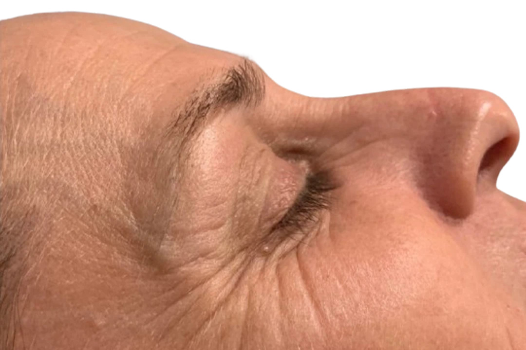
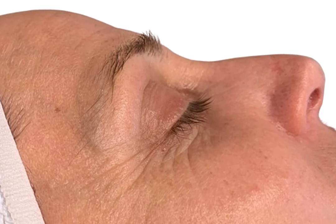
We have no before-and-after photographs of our own under-eye patients to publish yet, and we are not going to borrow one that looks like a dark-circle result when it is not. The one pair below is the only periorbital frame in the set Dermapen sent us for the DP4, the mechanical microneedling device described above. It shows lines at the outer corner of the eye. It is not a dark-circle result, it is not an under-eye bag result, and it says nothing about vascular discoloration, pigment or herniated fat. The full set is on the before and after page.
-
Lines fanning from the outer corner of the eye, with crepey skin across the temple. In the after photograph the fan of fine lines is shallower and less dense; the deeper crease beneath the eye is still visible. The head is turned slightly differently in the two frames.
Dermapen 4 marketing photographs · supplied by the manufacturer · no clinician credited · not a patient of this practice
Questions
Can laser get rid of my under-eye bags?
If the “bag” is shadow, discoloration, or crepey skin — yes, substantially. If it is herniated orbital fat, no. Those are different problems and the consultation exists to tell them apart.
I’ve been told my dark circles are genetic and untreatable. Is that true?
Partly. Constitutional pigment is genuinely stubborn and we will be honest about how much we can move it. But the vascular component — the blue-violet transmission through thin lid skin — is highly treatable and gets dismissed as “genetic” all the time. If your circles fade when you stretch the skin, you have been told the wrong thing.
Why does my under-eye look worse in some photos than others?
Lighting. Overhead or oblique light casts a shadow into the groove; flat frontal light erases it. If the difference between a ceiling light and a window is dramatic, your problem is structural shadow rather than pigment — and structural shadow is usually a cheek problem.
Do I need tear trough filler?
Probably less than you think. Most tear trough shadow is driven by loss of support in the cheek below it, and rebuilding that support from a deep, safer plane addresses the cause rather than camouflaging the shadow. We work through color and skin quality first, then reassess. A lot of patients never get to the point of needing anything injected at the rim.
How long until I see results?
Vascular: often within the first week or two. Texture from resurfacing: 2–4 weeks, continuing to 6 months. PRP: 4–6 months for full effect. Filler: immediate, settling over 2 weeks. Anyone quoting collagen results at one week is describing swelling.
Is this safe around the eyes?
Yes, with correct technique and the right eye protection for where we are working. At or inside the orbital rim we use internal metal corneal shields; nothing worn outside the eye protects it there. Outside the rim, external shielding is appropriate. Ask any provider you are considering which they use and where — if the answer is “goggles” for work on the lid, that protects them, not you.
What does it cost?
The consultation is free, as it is for everything here, and you leave with a written assessment whether or not you book anything. Vascular laser for a small area is $300, microneedling is $350 and RF microneedling is $750 for the face. Er:YAG resurfacing around the eye is not a separate line; it is priced as part of a full-face treatment. Midface filler is priced per syringe on the dermal fillers page.
Two ways to book
Call us
(757) 586-4575. Answered 24 hours a day, including evenings and Saturdays. Most people are booked in about a minute.
Book online
Pick your time on our scheduler — no account or login needed. When you're done, you'll land back here on a Booking confirmed page.
Didn't reach the confirmation page? Then nothing was booked — give us a call and we'll take care of it.
Newport News · Virginia
Find out what’s actually causing it.
The consultation is free. You leave with a written assessment of which causes you have, what we can treat, what we cannot, and what it costs — before you commit to anything.
Takes about a minute. You'll come back here to a confirmation page when you're booked — if you don't get there, call (757) 586-4575, any hour.